MCAT: Biology and biochemistry
Cells, genetics, metabolism, enzymes, amino acids, and organ systems explained together
On the exam: Cells, genetics, metabolism, enzymes, amino acids, the major organ systems. The largest share of science content.
Think biology on the MCAT is just memorizing organelle names? It's actually the biggest chunk of the science sections, and it rewards understanding over memorization. This video covers cells, genetics, metabolism, enzymes, amino acids, and the organ systems, then walks through a typical question step by step.
Cell Structure and Organelles
Picture a cell as a tiny factory. The nucleus holds the DNA blueprint. Mitochondria burn glucose and oxygen to make ATP, the cell's energy currency. Ribosomes on the rough endoplasmic reticulum build proteins, which the Golgi apparatus then folds, modifies, and ships to their destination. Lysosomes break down waste. Every organelle has one job, and matching structure to function is a classic question style.
Key points.
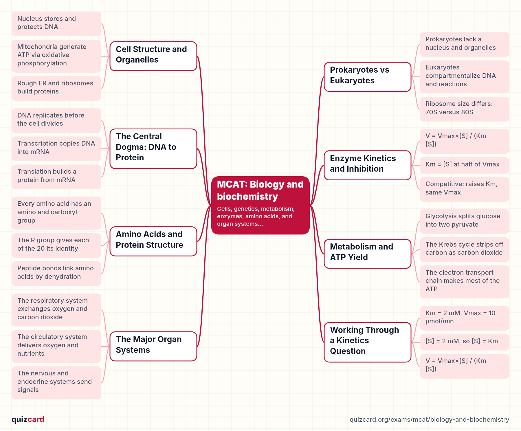
- Nucleus stores and protects DNA
- Mitochondria generate ATP via oxidative phosphorylation
- Rough ER and ribosomes build proteins
- Golgi apparatus modifies and ships proteins
Prokaryotes vs Eukaryotes
Bacteria are prokaryotes: their circular DNA floats free in the cytoplasm, and they have no membrane-bound organelles, just smaller seventy-S ribosomes. Animal and plant cells are eukaryotes: DNA is packaged as linear chromosomes inside a true nucleus, and specialized organelles like mitochondria and the endoplasmic reticulum compartmentalize reactions. The ribosome size difference matters clinically too, since some antibiotics target bacterial seventy-S ribosomes without touching our eighty-S ones.
Prokaryotic
- No nucleus, circular DNA
- No membrane-bound organelles
- 70S ribosomes
- Example: bacteria
Eukaryotic
- True nucleus, linear chromosomes
- Membrane-bound organelles present
- 80S ribosomes
- Example: animal and plant cells
The Central Dogma: DNA to Protein
The central dogma says information flows from DNA to RNA to protein. First, DNA replicates so each new cell gets a full copy. Transcription then copies one gene into messenger RNA inside the nucleus. That mRNA travels to a ribosome, where translation reads it three bases at a time, called a codon, to string together amino acids. Finally the chain folds and gets modified into a working protein, like insulin.
- DNA replicates before cell division
- Transcription copies DNA into messenger RNA
- Translation reads mRNA to build a protein
- Post-translational modification folds and finishes it
Enzyme Kinetics and Inhibition
Enzyme kinetics is described by the Michaelis-Menten equation: V equals Vmax times substrate concentration, divided by Km plus substrate concentration. Km is defined as the substrate concentration where the reaction runs at half of Vmax, and it tells you how tightly the enzyme binds its substrate. A competitive inhibitor fights for the active site, so it raises the apparent Km but leaves Vmax unchanged. A noncompetitive inhibitor binds elsewhere, leaving Km the same but dragging Vmax down.
V = Vmax×[S] / (Km + [S])
Km = [S] at half of Vmax
Competitive: raises Km, same Vmax
Noncompetitive: same Km, lowers Vmax
Key points.
- Enzymes speed reactions without being consumed
- Km reflects how tightly substrate binds
- Competitive inhibitors compete at the active site
- Noncompetitive inhibitors bind elsewhere and slow Vmax
Amino Acids and Protein Structure
All twenty standard amino acids share the same backbone: a central alpha carbon attached to an amino group, a carboxyl group, and a hydrogen, with a variable R group that makes each one unique, hydrophobic, acidic, basic, or polar. Amino acids link through peptide bonds, formed by dehydration synthesis, into a polypeptide chain. That chain folds through four levels of structure, primary sequence, secondary helices and sheets, tertiary three-dimensional shape, and quaternary, like the four subunits of hemoglobin.
Key points.
- Every amino acid has an amino and carboxyl group
- The R group gives each of the 20 its identity
- Peptide bonds link amino acids by dehydration
- Four levels build primary to quaternary structure
Metabolism and ATP Yield
One glucose molecule, fully broken down, nets roughly thirty-six ATP, though many modern estimates put it closer to thirty. Glycolysis splits glucose into two pyruvate molecules in the cytoplasm, yielding a small amount of ATP directly. Pyruvate then enters the mitochondria, feeding the Krebs cycle, which strips off carbon dioxide and loads electron carriers. Those carriers hand electrons to the electron transport chain, which uses oxygen as the final acceptor to generate the bulk of the ATP.
~36 ATP Net ATP produced per glucose through glycolysis, the Krebs cycle, and the electron transport chain
Key points.
- Glycolysis splits glucose into two pyruvate
- The Krebs cycle strips off carbon as carbon dioxide
- The electron transport chain makes most of the ATP
- Oxygen is the final electron acceptor
The Major Organ Systems
Each organ system supports the others. The respiratory system brings in oxygen and removes carbon dioxide. The circulatory system, powered by the heart, carries that oxygen and nutrients to every tissue. The nervous system sends fast electrical signals, while the endocrine system sends slower hormonal ones, and both rely on negative feedback loops, like insulin and glucagon balancing blood sugar, to keep the body's internal environment stable.
Key points.
- The respiratory system exchanges oxygen and carbon dioxide
- The circulatory system delivers oxygen and nutrients
- The nervous and endocrine systems send signals
- Negative feedback keeps the body in balance
Working Through a Kinetics Question
Here's a typical question. An enzyme has a Km of two millimolar and a Vmax of ten micromoles per minute. At a substrate concentration of two millimolar, what is the velocity? Notice the substrate concentration equals Km, which by definition means the reaction runs at half of Vmax. Plugging into the equation confirms it: ten times two, divided by two plus two, equals five micromoles per minute, half of Vmax, without needing a calculator.
Km = 2 mM, Vmax = 10 μmol/min
[S] = 2 mM, so [S] = Km
V = Vmax×[S] / (Km + [S])
V = (10×2)/(2+2) = 5 μmol/min
Key points.
- Question gives Km and Vmax for an enzyme
- Asks for reaction velocity at a given substrate level
- Plug values into the Michaelis-Menten equation
- Recognize substrate equal to Km means half-maximal velocity
Remember this
The one thing to remember: structure always explains function, from organelles to enzymes to organ systems. Quiz yourself on amino acid properties and enzyme kinetics before your MCAT test day, and the rest starts clicking into place.
Practice this unit
- Biology: Cell Structure and Function quiz18 multiple-choice questions with explanations · no account needed
- AP Biology: Genetics and Heredity quiz18 multiple-choice questions with explanations · no account needed
- Chemistry: Atomic Structure and Periodic Trends quiz13 multiple-choice questions with explanations · no account needed
- Biology: Cell Structure and Function30 flashcards · study free
- AP Biology: Genetics and Heredity30 flashcards · study free
- Organic Chemistry: Functional Groups and Reactions30 flashcards · study free
- Chemistry: Stoichiometry and Chemical Reactions30 flashcards · study free
Every MCAT deck and quiz: the MCAT hub.
Other MCAT unit guides
- CARSStudy guide
- General chemistryStudy guide
- Organic chemistryStudy guide
- PhysicsStudy guide
- Psychology and sociologyStudy guide
Try it with your own material
Upload a PDF, slides or notes and study the deck in minutes. Making your own cards is free with no limits.